Home
/ Anatomy Of The Back Of The Neck Muscles - Cervical Motor Control Part 1 Clinical Anatomy Of Cervical Spine Rayner Smale _ And the activity at our anterior neck muscles is connected in a wonderful way to the activity of.
Anatomy Of The Back Of The Neck Muscles - Cervical Motor Control Part 1 Clinical Anatomy Of Cervical Spine Rayner Smale _ And the activity at our anterior neck muscles is connected in a wonderful way to the activity of.
Anatomy Of The Back Of The Neck Muscles - Cervical Motor Control Part 1 Clinical Anatomy Of Cervical Spine Rayner Smale _ And the activity at our anterior neck muscles is connected in a wonderful way to the activity of.. The neck muscles, including the sternocleidomastoid and the trapezius, are responsible for the gross motor movement in the muscular system of the head and neck. Indeed it is rare to find a client/patient whose neck is not tight. Muscles of the back can be divided into superficial, intermediate, and deep group. The thyrohyoid is a quadrilateral muscle located in the muscular triangle of the neck. The back anatomy includes some of the most massive and functionally important muscles in the human body.
The extensors and rotators of the head and neck: Rectus capitis posterior major and rectus capitis posterior minor attach the inferior nuchal line of the occiput to the c2 and c1 vertebrae respectively. Table of contents the extrinsic muscles of the back: Intermediate back muscles and c. The ligamentum nuchae separates the muscles of the two sides of neck.
Muscles Move And Support The Spine from www.spineuniverse.com The classic computer position shortens the posterior (back) neck muscles, making them tight and, over time, possibly shorter. Its attachment to the frontal and occipital bellies (muscles on the brow at the front and on the upper back of the head). This article provides an overview of the neck muscles, their anatomy, origins, insertions, actions, and innervation. Instead, i think it's simply better to break movements down into three categories Tutorials and quizzes on the anatomy and actions of the back muscles (iliocostalis, longissimus, spinalis, multifidus, and quadratus lumborum), using interactive animations, diagrams, and illustrations. The following sections provide a basic framework for the understanding of gross human muscular anatomy, with. Explanations spinoscapular and spinohumeral muscles the extrinsic back muscles are also referred to as secondary back muscles. I have covered a complete anatomy of the back muscles, explaining the important muscles, their locations and functions.
Injuries of the intrinsic back muscles often occur while using improper lifting technique.
The sternocleidomastoid muscle, or scm, is one of the larger and more superficial muscles in the neck, making it an important and easily identifiable anatomical landmark. Click now to learn more at kenhub! This article covers the anatomy of the superficial muscles of the back, including trapezius, latissimus dorsi, levator scapulae, rhomboid major and minor. Learn about anatomy neck muscles with free interactive flashcards. Other muscles in the back are associated with the movement of the neck and shoulders. Its attachment to the frontal and occipital bellies (muscles on the brow at the front and on the upper back of the head). The extensors and rotators of the head and neck: Injuries of the intrinsic back muscles often occur while using improper lifting technique. Several other muscles of the back also extend up to the neck region and are partly connected with the cervical part of the vertebral column, including the trapezius, levator scapulae, splenius, iliocostalis, longissimus, rotatores, semispinalis, interspinales, and intertransversarii muscles. Muscles of the head & neck | anatomy model. Beneath the integument the back of neck presents in the median plane the ligamentum nuchae, which is a triangular fibrous sheet and represents upward continuation of supraspinous ligament. Educational video describing the muscle anatomy of the neck. The neck muscles are specifically designed to either allow for neck movement or to provide structural support for the head.
The classic computer position shortens the posterior (back) neck muscles, making them tight and, over time, possibly shorter. I have covered a complete anatomy of the back muscles, explaining the important muscles, their locations and functions. The superficial back muscles are covered by skin, subcutaneous connective tissue and a layer of fat. The surface muscles of the upper back include the trapezius muscles (traps) and posterior deltoids. Accordingly, the anterior (front) neck muscles can become long and weak.
Levator Scapula Muscle And Its Role In Pain And Posture from www.verywellhealth.com Four groups of muscles in neck. Its attachment to the frontal and occipital bellies (muscles on the brow at the front and on the upper back of the head). Accordingly, the anterior (front) neck muscles can become long and weak. The muscles of the back that work together to support the spine, help keep the body upright and allow twist and bend in many directions. I have covered a complete anatomy of the back muscles, explaining the important muscles, their locations and functions. The splenius muscles originate at the midline and run laterally and superiorly to their insertions. Memorize all the muscle facts with the help of muscle cheat sheets. In fact, the back contains a group of muscles, not one muscle.
Muscle spasming of the neck is likely the most common musculoskeletal complaint that exists.
The splenius muscles originate at the midline and run laterally and superiorly to their insertions. If we want to locate the back muscles in the body, we can say that it starts from the top of the neck and ends. The splenius muscles originate at the midline and run laterally and superiorly to their insertions. The trapezius muscle actually considered to be just as much of a muscle related to the back as it is the neck. Several other muscles of the back also extend up to the neck region and are partly connected with the cervical part of the vertebral column, including the trapezius, levator scapulae, splenius, iliocostalis, longissimus, rotatores, semispinalis, interspinales, and intertransversarii muscles. The ligamentum nuchae separates the muscles of the two sides of neck. I have covered a complete anatomy of the back muscles, explaining the important muscles, their locations and functions. Obliquus capitis superior also extends from the occiput to c1 while obliquus. The physicians originally studying human anatomy thought the skull looked like an apple. From the sides and the back of the neck, the splenius capitis inserts onto the head region, and the splenius. Table of contents the extrinsic muscles of the back: There are a number of specific muscles within the back anatomy, and its important to take a quick look at all of them to see how you can target them efficiently and develop a strong back. The back muscles can be three types.
The trapezius muscle can be involved in extending the head upward or neck backward. The splenius capitis and cervicis (spinotransversales muscles). Beneath the integument the back of neck presents in the median plane the ligamentum nuchae, which is a triangular fibrous sheet and represents upward continuation of supraspinous ligament. The sternocleidomastoid muscle, or scm, is one of the larger and more superficial muscles in the neck, making it an important and easily identifiable anatomical landmark. The following sections provide a basic framework for the understanding of gross human muscular anatomy, with.
Muscles Of The Head And Neck Anatomy Pictures And Information from innerbody.imgix.net Memorize all the muscle facts with the help of muscle cheat sheets. These muscles give height and breadth to back development. Muscle spasming of the neck is likely the most common musculoskeletal complaint that exists. The surface muscles of the upper back include the trapezius muscles (traps) and posterior deltoids. Accordingly, the anterior (front) neck muscles can become long and weak. Other muscles in the back are associated with the movement of the neck and shoulders. Working in pairs on the. The extensors and rotators of the head and neck:
Intermediate back muscles and c.
The muscles in all of the layers are innervated by the posterior rami of spinal nerves: The extensors and rotators of the head and neck: In this anatomy course, part of the anatomy specialization, you will learn how the components of the integumentary system help protect our body (epidermis, dermis, hair, nails, and glands), and how the musculoskeletal system (bones, joints, and skeletal muscles) protects and allows the body to move. Its attachment to the frontal and occipital bellies (muscles on the brow at the front and on the upper back of the head). The splenius capitis and cervicis (spinotransversales muscles). Four groups of muscles in neck. Educational video describing the muscle anatomy of the neck. The neck is the part of the body on many vertebrates that connects the head with the torso and provides the mobility and movements of the head. The surface muscles of the upper back include the trapezius muscles (traps) and posterior deltoids. The classic computer position shortens the posterior (back) neck muscles, making them tight and, over time, possibly shorter. Memorize all the muscle facts with the help of muscle cheat sheets. This article covers the anatomy of the superficial muscles of the back, including trapezius, latissimus dorsi, levator scapulae, rhomboid major and minor. From the sides and the back of the neck, the splenius capitis inserts onto the head region, and the splenius.
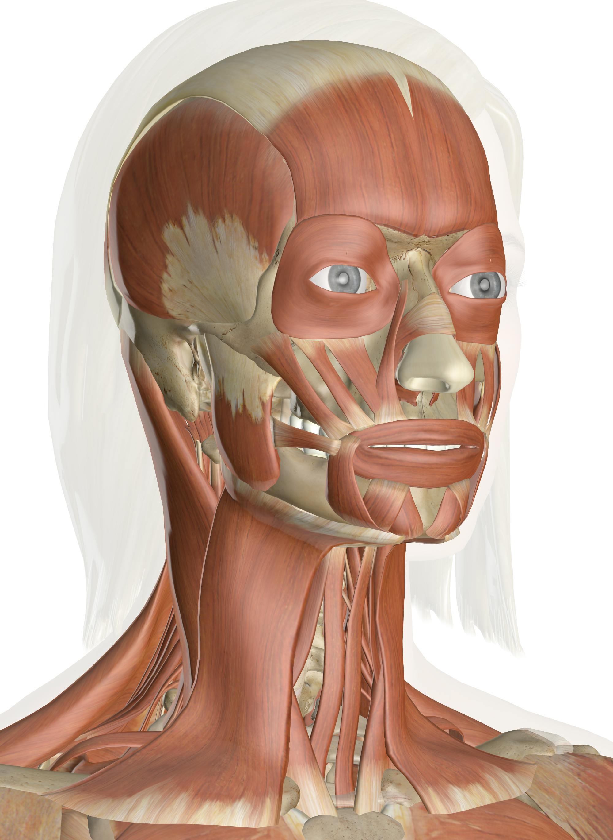
Explanations spinoscapular and spinohumeral muscles the extrinsic back muscles are also referred to as secondary back muscles anatomy of back of neck. The image below to shows all the significant back muscles (in addition to some neck muscles)

:max_bytes(150000):strip_icc()/GettyImages-499158129-56a05f075f9b58eba4b0267f.jpg)